
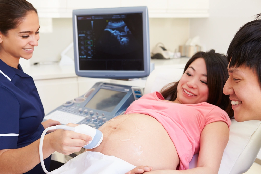
Every expectant mother looks forward to that first ultrasound image when they get to see their little one hanging out in their belly. Today, most mothers might not be able to imagine going through an entire pregnancy without this amazing technology. And yet, mothers before the 1970s mostly did. So what’s the story behind this technology, and how did it become a standard operating procedure for monitoring pregnancy?
The origin of the technology may surprise you. Let’s take a look.
ULTRASOUND TECHNOLOGY EMERGED FROM BATS & A WORLD AT WAR
The field of radiology has a long and complicated history. The story begins with echolocation and a furry little flying creature that doesn’t see very well. The study of bats and ultrasonic ranging was monumental. Bats use high-frequency sounds to detect motion and objects. So it was thanks to bats the concept of ultrasonic technology emerged and could be further explored during the war.
Like many technological innovations, the concept and tech behind ultrasound were born out of necessity in wartime. During World War I, the French government wanted the creation of a device that was capable of detecting enemy submarines submerged deep in the water. The development of such a device was put in the hands of Paul Langevin, who worked on it throughout the war. His tech, however, was not finished in time to help with the war effort, but the ground had been laid for ultrasonic devices and sonar detection.
The sinking of the Titanic just a few years earlier had also sparked interest in the development of ultrasonic technology. Ships needed better technology to navigate the dark waters and detect objects underneath (such as icebergs). People were also exceedingly curious about diving deep in the ocean and having visibility or some ability to detect objects. The hydrophone was invented in 1916, and it was the first iteration of the transducer.
Meanwhile, during the late 19th and 20th centuries, physicians were searching for ways to look inside the body in the medical field. Several probes and scopes for diagnostic purposes had been invented and were inching their way there, but the use of ultrasound for medical purposes was not put into play until the 1920s and 1930s.
The Use of Ultrasound in the Medical Field Begins
Sonography began its foray into the world of medicine through unexpected avenues. This technology was first used for physical therapy to help European soccer players heal. In the 1940s, ultrasound exploded in the medical field and became the go-to for many diagnostic scenarios. Sonography would also be used for arthritic pain in stomach ulcers.
The Fetal Ultrasound Breaks Through
Ultrasound technology started sprouting up in maternity clinics in the 1950s in Scotland and England. The Scots began to use it with some frequency, and the procedure started taking off in England a few years later. In the 60s and 70s, what is known as the sonic boom hit the medical community in the United States by storm with the invention of the Doppler, which allowed the detection of blood flow.
Towards the final decades of the 20th century, fetal ultrasound became routine in maternity clinics.
How Does It Work?
Ultrasound imaging works by bouncing back ‘ultrasonic’ waves into human tissue or structure. The waves are above the audible range of human hearing, but they produce images as they bounce back through the transducer. This transducer converts the sound waves into an electronic image.
WHAT CAN IT TELL YOU ABOUT YOUR BABY?
The fetal ultrasound is a routine part of prenatal care. The procedure gives physicians a lot of information about your developing baby, which is considered very low risk. The ultrasound for your baby includes checking for:
- Normal development
- Head and brain development
- Development of other body parts
- Kidneys
- The placement of the placenta
- Spine abnormalities
- Bladder
The ultrasound can also help with:
- Showing if you are expecting twins (or multiple babies)
- Indicating the gestational age
- Showing the growth of the baby
There is a growing use of ultrasound in detecting fetal abnormalities and genetic syndromes. Today, 3-D ultrasounds can increase the clarity of suspected problems and may be ordered if a 2-D image suggests something abnormal. However, there is still a risk for false positives (detecting something that is not there) or false negatives (not detecting something that is there). The technology is not foolproof.
VISIT YOUR FAMILY DOCTOR FOR FURTHER QUESTIONS ABOUT YOU AND YOUR BABY’S HEALTH
Expecting a new member of the family is an exciting time. It can also be overwhelming, stressful, and scary. The good news is that the world of medicine has seen incredible advancements in technology in every direction. EP Family & Ped stays close to expecting mothers as they await the big day as a family medicine practice. Having a trusted primary care physician means there is someone in your corner when questions arise. We like to think we are part of the team, along with your gynecologist and other medical professionals that form part of prenatal care.
Your primary care physician is there before, during, and after. El Paso Family & Pediatric Clinic sees entire families and practices primary care that sees the family as a unit, offering pediatric care as well as your little one grows.
Want to find a family medicine clinic that offers quality care in El Paso? Call EP Family & Ped today and learn about our services.